All Facilities | All Instruments
MALDI Imaging (Tissue Histology) Facility

The MALDI Imaging (Tissue Histology) Facility at the CUNY ASRC is the first core facility with MALDI-TOF MS imaging capacity in the New York metropolitan area. The facility provides MALDI Imaging services to research scientists including tissue preparation, matrix coating, MALDI-MS profiling, and imaging/data analysis. MALDI-MS Imaging is a powerful tool for label-free in situ profiling of proteins, lipids, metabolites and small molecules. Both research institutions and industrial enterprises are welcome to utilize our facility.
The MALDI-TOF MS Imaging Facility is jointly overseen by the Structural Biology Initiative and Neuroscience Initiative.
Complete the online application to become a user of this and other ASRC facilities.
Facility Details
-
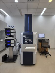
Bruker timsTOF-flex MALDI-2 with microGRID ›
The Bruker timsTOF fleX equipped with MALDI-2 and microGRID technology provides advanced capabilities for high-resolution MALDI imaging of biological tissues. MALDI-2 post-ionization significantly enhances ion yields, particularly for low-abundance and poorly ionizing molecules such as lipids and metabolites, thereby improving sensitivity and molecular coverage. The microGRID feature enables precise, reproducible laser positioning and finely controlled sampling patterns, supporting high spatial resolution and uniform pixel-to-pixel data quality across large tissue areas. Together, these capabilities allow for detailed mapping of molecular distributions with improved dynamic range and reproducibility. As a result, the system is well suited for spatial metabolomics and lipidomics studies requiring both high sensitivity and spatial fidelity.
-
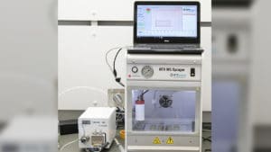
HTX M5 Matrix Sprayer ›
The HTX M5 Matrix Sprayer is an automated system designed to deliver highly uniform and reproducible matrix coatings for MALDI imaging experiments. It utilizes controlled pneumatic spraying and programmable parameters to generate fine, consistent droplets, ensuring homogeneous matrix crystallization across tissue sections. This level of control minimizes analyte delocalization while enhancing signal intensity and reproducibility, which is critical for high-quality spatial analysis. The system supports a wide range of matrices and application conditions, making it well suited for both metabolomic and proteomic MALDI imaging workflows.
-
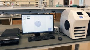
Leica Aperio CS2 Slide Scanner ›
The Leica Aperio CS2 is a high-throughput digital pathology system designed for rapid, high-resolution scanning of whole tissue slides. It produces brightfield images with excellent color fidelity and detail, enabling accurate visualization of tissue morphology and histological features. The scanner supports efficient digitization and archiving of large slide collections, facilitating downstream analysis and data sharing. In workflows such as MALDI imaging, these high-quality optical images provide essential spatial context for correlating molecular distributions with histological structures.
-
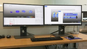
SCiLS MALDI Imaging Analysis Workstation ›
The SCiLS Lab is a specialized platform for the visualization, processing, and statistical analysis of MALDI imaging mass spectrometry data. It enables users to explore spatial distributions of molecular features across tissue sections through intuitive image rendering and segmentation tools. The software incorporates advanced statistical methods, including multivariate analysis and machine learning, to identify discriminative molecular patterns and regions of interest. Additionally, SCiLS Lab supports integration with external databases and annotation tools, facilitating more confident interpretation of complex imaging datasets.


All MSI Services require prior consultation with the core for estimated cost and timeline.
Every MSI project is unique – there is no standard method that will work for any sample. The instruments used, sample preparation, data acquisition method, and analysis software will depend on the tissue type and size, the compounds of interest, the spatial resolution needed, and the goals of each experiment. Additional experiments to assist with or complement MSI may be needed, for example, LC-MS analysis and/or microscopy.
Please contact us to inquire about your project.
For more information, please contact:
-
Ye He, Ph.D.
- Director, Live Imaging and Bioenergetics Facility
- Co-Director, MALDI-TOF MS Imaging Facility
- Research Associate Professor, Neuroscience Initiative
Phone: 212.413.3182
-
Rinat Abzalimov, Ph.D.
- Director, Mass Spectrometry Facility
- Co-Director, MALDI-TOF MS Imaging Facility
- Research Associate Professor, Structural Biology Initiative
Phone: 212.413.3236
