Posted on November 23, 2020 in ASRC News, Nanoscience Initiative
When scientists design a novel material, they sometimes don’t understand all of its properties. To use the material to its fullest potential, they first need to see how it works in action.
Now, a study from the Advanced Science Research Center at The Graduate Center, CUNY (CUNY ASRC) demonstrates a new way to visualize nanostructures made of short peptide sequences. Not only does the method allow for very high resolution, but it also lets scientists watch how these bio-inspired soft materials — which have applications in health care, food, and green energy — behave in real time, and in situ.

The work, published in ACS Nano, comes from the lab of Rein Ulijn, director of the Nanoscience Initiative at CUNY ASRC and Albert Einstein Professor of Chemistry at The Graduate Center and Hunter College.
“Our design demonstrates that peptide nanofibers, which are protein-like structures, can be easily visualized with super-resolution microscopy to monitor a metabolic process in real-time,” said Mohit Kumar, lead author on the paper and a research associate in the Ulijn lab. “Apart from the soft material research community, it has the potential to greatly impact the biomedical field and health care industry.”
Key to their method, the researchers found a new way to label the peptides with dye for fluorescence microscopy. Dyes are usually attached via a strong chemical bond, but this is tedious and can affect how the material behaves, Kumar explained. Instead, the team attached their dye with weaker, electrostatic forces that come from the attraction between positive and negative charges.
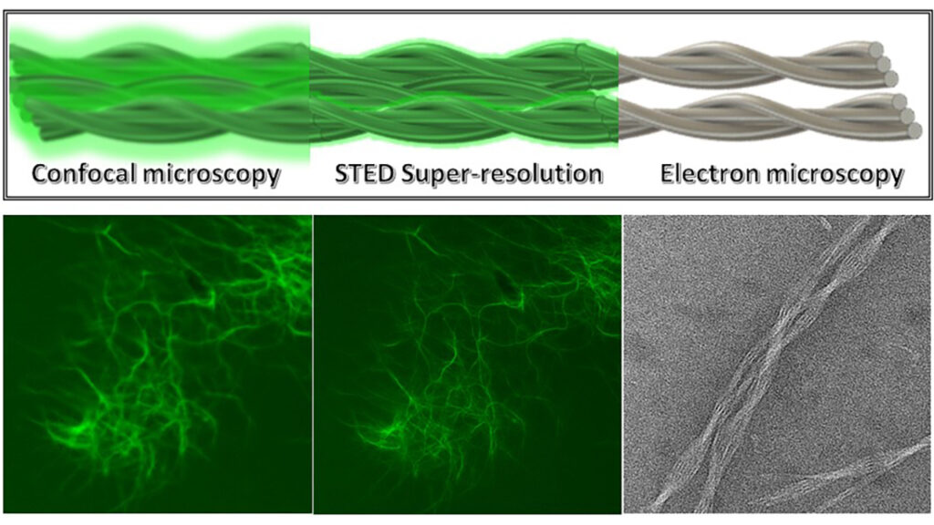
Combining this method with stimulated emission depletion microscopy, they obtained high-resolution images and were able to watch a live video of the nanofibers being degraded by enzymatic hydrolysis.
Their work overcomes limitations of other techniques. Electron microscopy, for instance, has high resolution but the sample needs to be dry. An optical microscope, on the other hand, allows for in situ observation but has lower resolution.
The next step will be to test the method with biological samples like cells and tissues, rather than with synthetic materials, so that it can someday be used for in vivo models.
