Several pieces of equipment in this facility provide an integrated set of different biophysical approaches for studies of macromolecular structure and interactions. The below instrumentation is available as of 2024.
For further information, please contact Structural Biology Initiative Director Kevin Gardner at kgardner@gc.cuny.edu or via telephone at 212.413.3220 for further information.

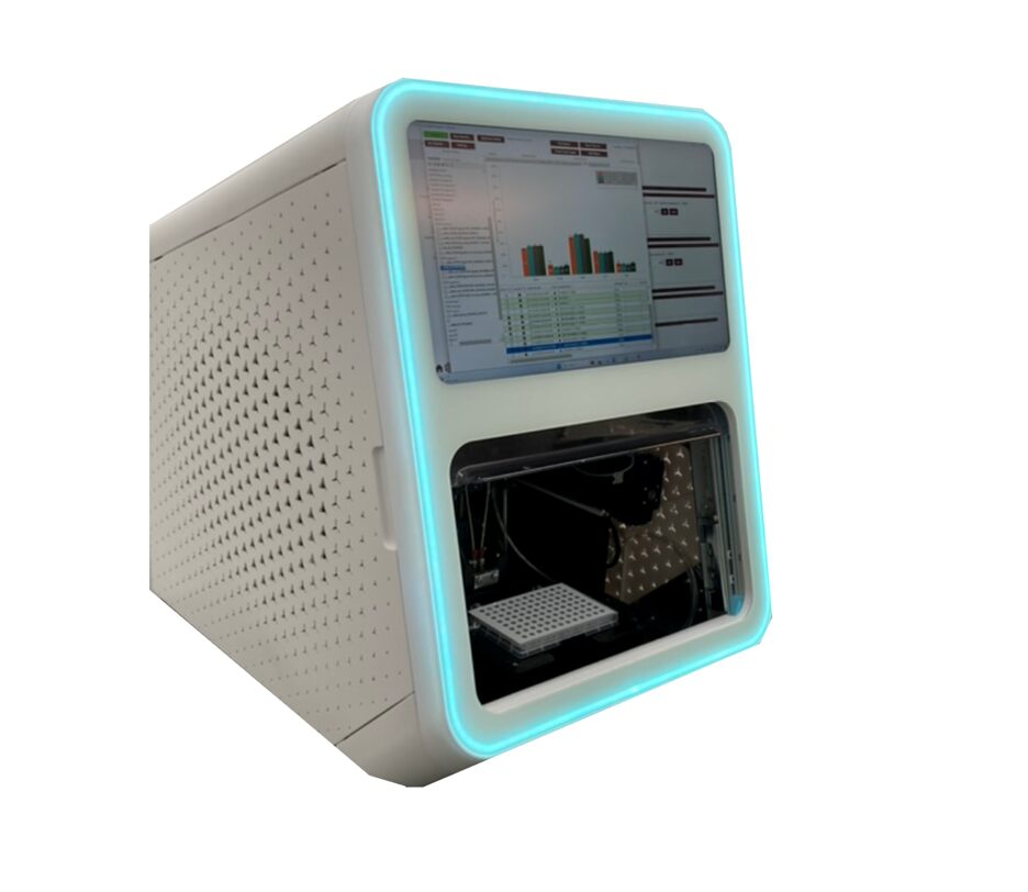
Using microfluidic modulated spectroscopy, the Aurora offers automated infrared spectroscopy measurements with real-time buffer subtraction. Focusing on the Amide I band, changes in secondary structure elements can be analyzed after ligand binding, mutagenesis, or denaturing treatments. Advantages include 96-well plate automation, wide range of buffer compatibilities, and easy to use software for running experiments and processing data.

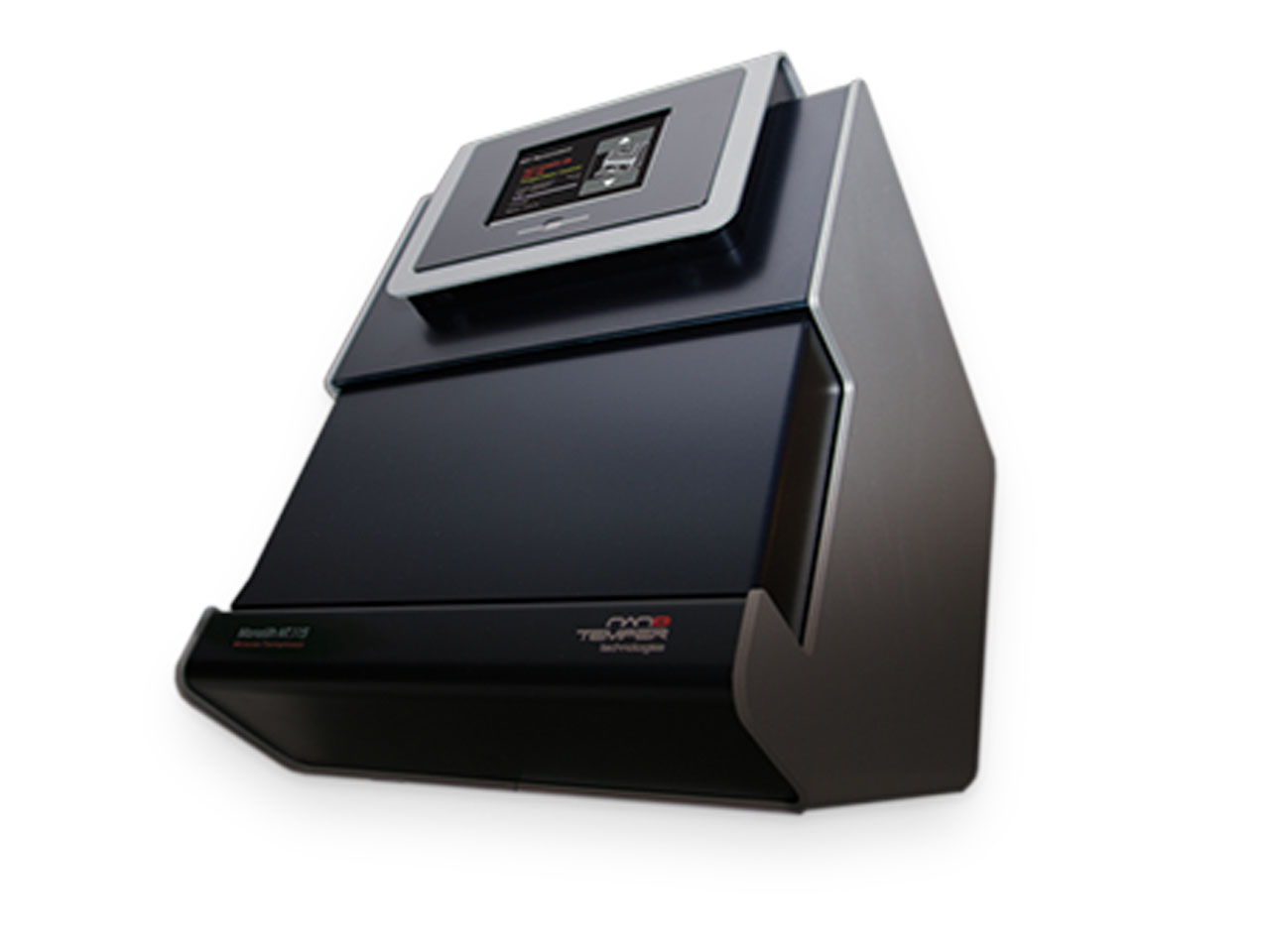
This instrument uses changes in thermophoretic mobility – the migration of macromolecules into or away from a zone with elevated temperature – to characterize binding reactions. Advantages of this instrument include low sample consumption (ca. 100 microliters of low micromolar protein samples) and speed of measurement (under one hour for 16 point titration curve).

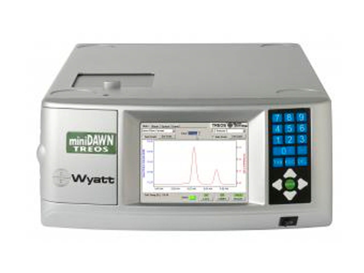
Provides independent measurements of molar mass and size of macromolecules in solution, particularly when coupled with Superdex 75 or Superdex 200 gel filtration chromatography (SEC-MALS).

(instrument transferred to Nanoscience Initiative, Spring 2024; contact us for information on use)
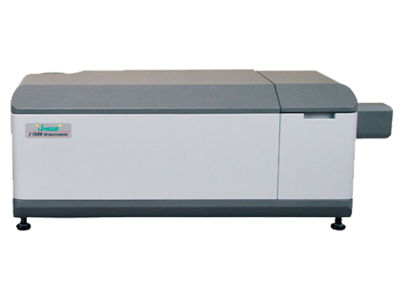
Utilized for studies of protein secondary and tertiary structure, this spectrometer can detect changes in either circular dichroism (monitoring protein backbone environments) or fluorescence (monitoring Trp sidechains and other fluorescent groups). Accessories allow variable temperature operation, automated titrations and stopped-flow experiments.
