Given the importance of integrating functional studies – both in vitro and cell-based – into structural biology work, the Structural Biology Initiative includes equipment for a range of such studies.
For further information, please contact Kevin Gardner at kgardner@gc.cuny.edu or at 212.413.3220.

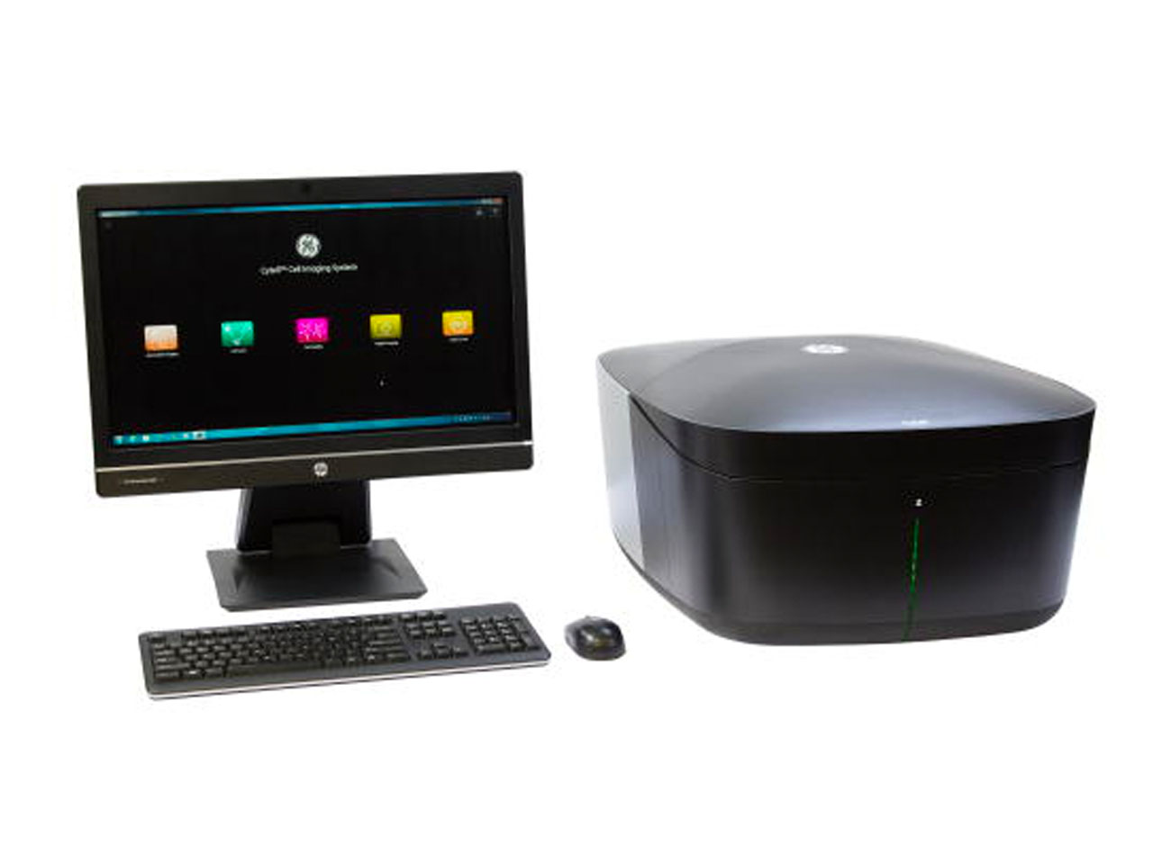
Simple automated cell microscopy instrument, allowing the straightforward collection of multicolor cellular images along with a number of cell-based assays with fluorescent or visible readouts, including assays of cell viability, cell cycle staging, gene expression and others.

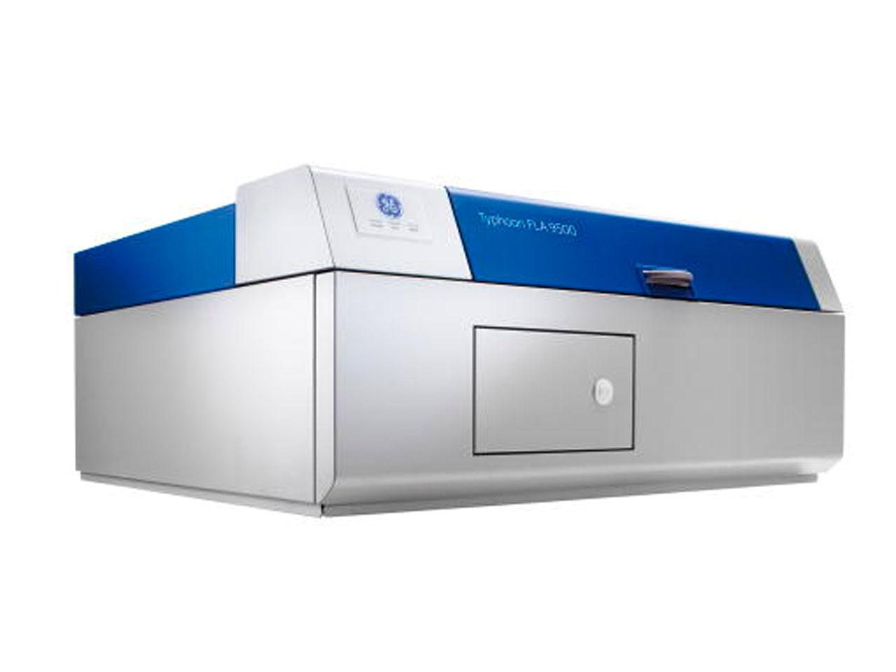
Designed to provide quantitative imaging of gels, blots and other two-dimensional formats of radiolabeled (32P) and fluorescently-labeled samples.

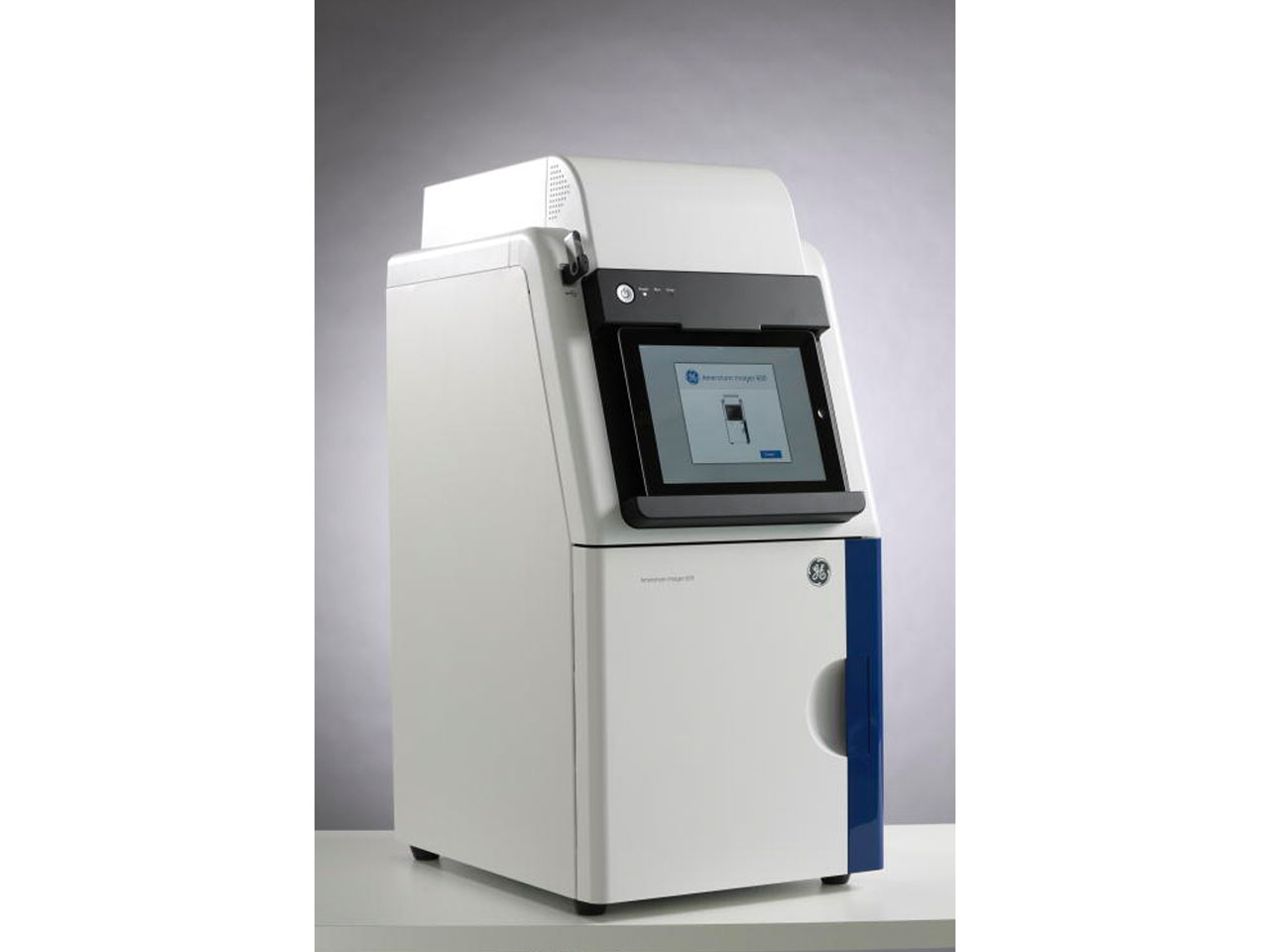
Used to image DNA gels (UV) and Western blots (chemiluminescence), but can image fluorescence as well.

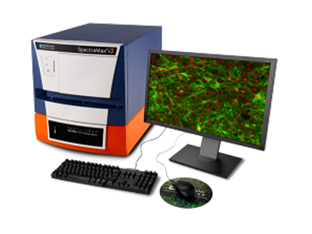
Supports a variety of plate-based biochemical assays, including those utilizing changes in absorbance, fluorescence, or luminescence. Additional capabilities to monitor changes in fluorescence polarization (FP) and AlphaScreen type assays are also equipped.
Cell Culture Facility
This instrument uses changes in thermophoretic mobility – the migration of macromolecules into or away from a zone with elevated temperature – to characterize binding reactions. Advantages of this instrument include low sample consumption (ca. 100 microliters of low micromolar protein samples) and speed of measurement (under one hour for 16 point titration curve).
