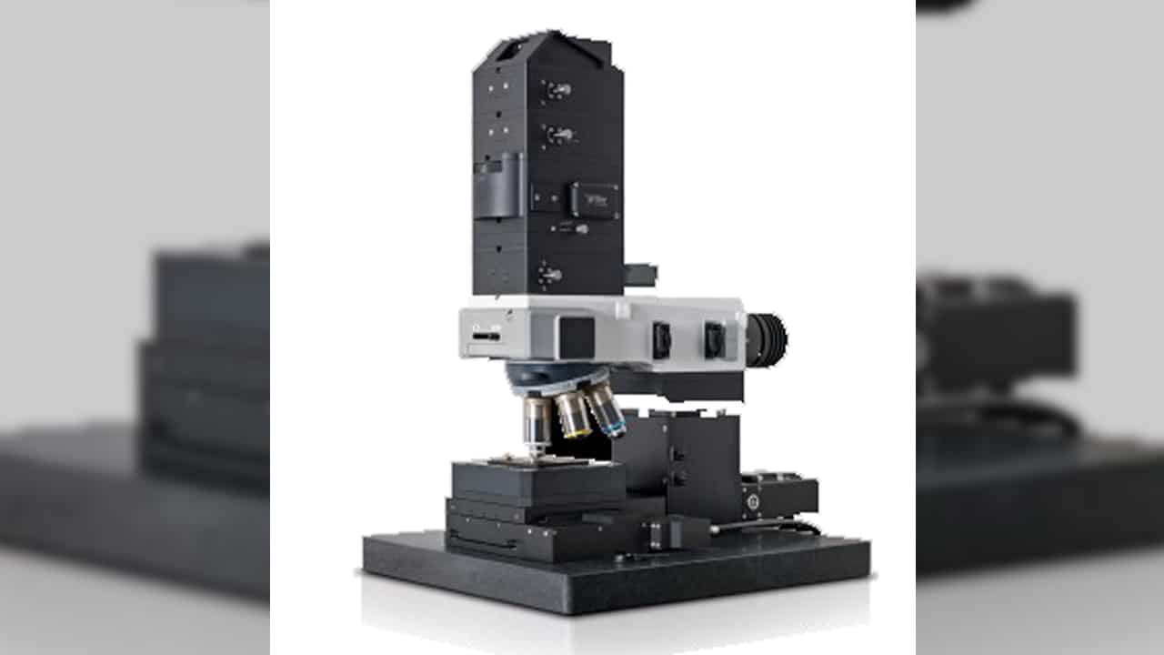
WITec Confocal Raman Microscope alpha300R

Raman imaging is non-destructive imaging techniques, no staining of fixation of sample is required. Raman imaging generates hyperspectral images with the information of a complete Raman spectrum at every image pixel. WITec alpha300R confocal Raman microscope enables confocal Raman imaging (both surface-scan and depth-scan) with excellent resolution and capability of time series. The ultra-high throughput spectroscopic system guarantees high sensitivity and best performance in spectral resolution.
Features
- Raman spectral imaging: acquisition of a complete Raman spectrum at every image pixel
- Planar (x-y-direction) and depth scans (z-direction) with manual sample positioning, time series
- Automated, motorized sample positioning and measuring with piezo-driven scan stages
- Additional Temperature control stage
- Single point Raman spectrum acquisition and single-point depth profiling
- Fibre-coupled UHTS spectrometer specifically designed for Raman microsopy and applications with low light intensities
- Bright/Dark Field Microscopy
- Two lasers: 532 nm and 633 nm.
- Five objective Lens for dry samples:
- 10X: Zeiss EC Epiplan-Neofluar, NA 0.25 DIC
- 50X: Zeiss EC Epiplan, NA 0.75 HD (darkfield only)
- 50X: Zeiss EC Epiplan-Neofluar, NA 0.8 DIC
- 100X: Zeiss EC Epiplan-Neofluar, NA 0.9 DIC
- 50X: Nikon DF Plan ELWD, NA 0.55 (heating stage only)
Contacts
- Tong Wang, Ph.D.
Manager, Imaging Suite
Research Associate Professor, Nanoscience Initiative
twang1@gc.cuny.edu
