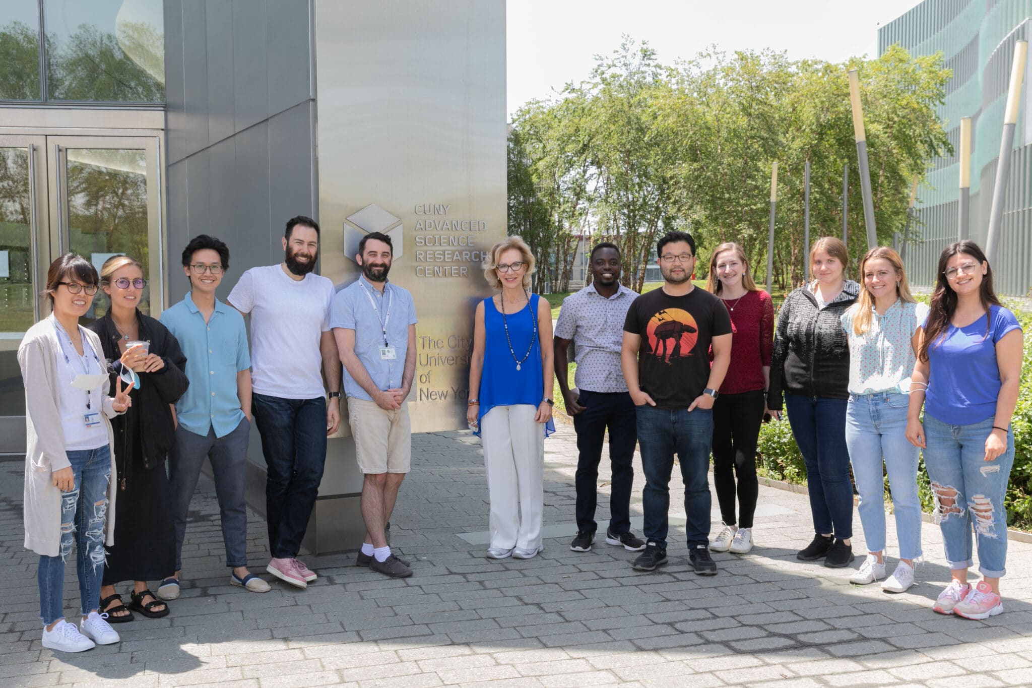
Professor Patrizia Casaccia, M.D., Ph.D., and her team use molecular and cellular techniques to understand how glial cells acquire their identity and respond to external stimuli. The team is interested in understanding how experiences (including social context and stress), dietary changes (metabolites and microbiota), and small changes in the brain microenvironment affect gene expression in the developing and adult brain. The methodologies used include molecular and robotic technologies for the study of human and rodent samples, dietary manipulations, study of metabolites, and behavioral assessment in models of neurological disorders. The team has a strong interest in defining mechanisms of neurodegeneration and brain tumors in order to develop novel therapeutics.
Team I

Define the molecular mechanisms responsible for acquisition and loss of oligodendrocyte cell identity
This area is related to understanding:
- How myelin is formed during development and during myelin repair and how the process is affected over time. The emphasis is on the definition of nuclear changes that result in the transcriptional program of oligodendrocyte progenitor differentiation in response to chemical and physical stimuli;
- How glial progenitors lose their “good” identity and may turn “bad” and form brain tumors.
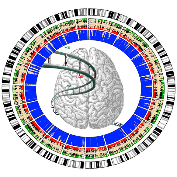
Figure: The concentric colored circles define chromosomal locations and the genome-wide distribution of differentially methylated genes (either hypermethylated or hypomethylated). We use epigenome-wide approaches to understand how cells reorganize their entire genetic information during the transition from progenitors to myelin-forming oligodendrocytes
Team II

Address mechanisms of neurodegeneration and mechanisms of disease progression in demyelinating disorders (e.g., Multiple Sclerosis)
This area is related to understanding the study of:
- Mitochondrial Bioenergetics studied in rat neurons exposed to the cerebrospinal fluid of human patients with different degrees of disability
- Study the molecular composition of the CSF to identify molecular pathways to be targeted to treat neurodegeneration
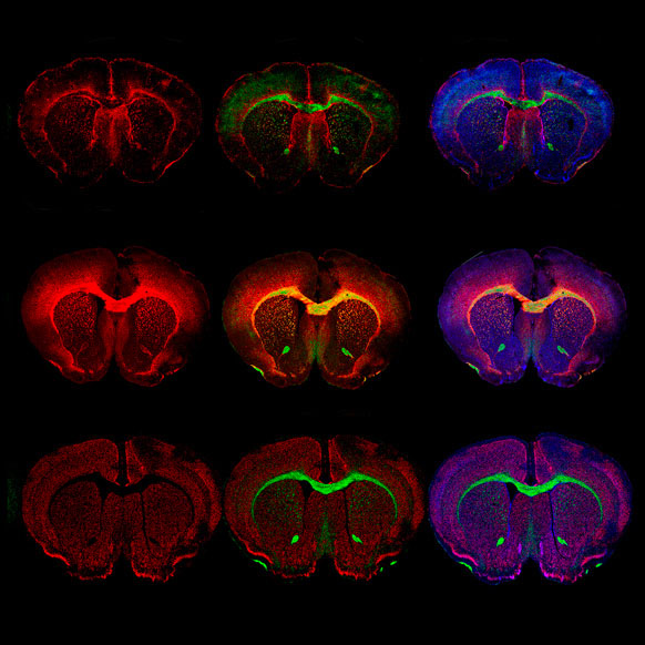
Figure: Serial coronal sections of adult mouse brains stained with fluorescent probes identifying myelinated fibers (in green and red) and all cell nuclei (blue).
Team III

Define whether and how the macro-environment (i.e., social experience; physical environment; diet) impacts development of the brain and susceptibility to disease
This area is related to understanding:
- The study of genome wide changes in DNA methylation and histone modifications using chromatin immunoprecipitation;
- Single cell transcriptomics;
- The study of gut/brain interaction: a two-way communication;
- The effect of diet on myelin and disease course;
- The effect of environmental variables on myelin formation and behavior.
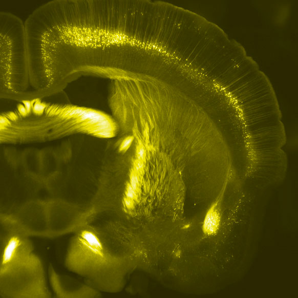
Figure: Coronal section of the brain of a transgenic mouse with green fluorescent protein expressed in selected neuronal populations.
